 In the early 20th Century, not long after X-rays were discovered, medical professionals recognized their value as diagnostic tools: They could clearly reveal structures hidden inside the body without the need for risky surgery. At the dawn of the 21st century, a revolutionary new technology has entered the diagnostic arena. Today, Cone Beam Computed Tomography (CBCT) promises to change the way many dental problems are diagnosed and treated.
Cone Beam CT has some similarities with conventional X-rays, and also with the standard CT scans you would get in a hospital setting. But it’s a quantum leap forward in technology and diagnostic precision. For the dentist, it offers the ability to visualize intricate structures inside the mouth, such as root canals, nerves and sinuses (air-filled spaces) in the jaw — in three dimensions — without surgery. For the patient, it can reduce the need for invasive procedures, shorten treatment time and offer the chance for a better outcome.
The detailed diagnostic images that CBCT provides have made it an essential tool in many dental specialties. But, as with any diagnostic tool that uses radiation, the medical benefits offered must be weighed against the (small) potential risks of the procedure.
How Cone Beam CT Works
X-rays, like visible light, are a form of energy on the electromagnetic spectrum. Just as light makes an image on photographic film (or a digital camera sensor), X-rays can also form an image. The difference is that energetic X-rays can penetrate bone and soft tissue, and reveal its hidden structure by their varying degrees of absorption; in other words, they form a grayscale picture of what’s underneath the surface. But conventional X-rays are limited: Like a still-life picture, they show only one perspective on the scene.
Now imagine a “flip book” — the kind of small book made up of a series of pictures, each slightly different. When you rapidly page through it, you may see (for example) an animated cartoon or a still subject from different perspectives. If you could put together a flip book made from a series of X-ray “slices” of the same subject, taken at slightly different angles, you would be able to create an “animation” of the X-rays. And from there, it’s only one more step to making a 3-D model.
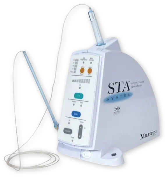
That’s exactly what CBCT scanners do. Using a rotating imaging device that moves around the patient’s head, the scanner records between 150 and 600 different X-ray views in under a minute. Then, a powerful computer processes the information and creates a virtual model of the area under study. When it’s done, the model appears as a three-dimensional image on a computer screen: It can be rotated from side to side or up and down, examined in greater or less detail, and manipulated in any number of ways — all without the patient feeling any discomfort… or even being present.
Where Cone Beam CT Is Used
The ability to see fine anatomical structures in 3-D has proven invaluable in treating conditions in many areas of dentistry.
- Orthodontics: Having accurate information on the position of teeth and jaws helps determine exactly how and where teeth should be moved.
- Dental implants: Detailed CBCT images are used to determine the optimum location for the titanium implants while avoiding nerves, sinuses and areas of low bone density.
- Orthognathic Jaw Surgery and Temporo-mandibular Joint (TMJ) Disease: Patients benefit when the specialists who treat these conditions can evaluate their anatomy with the three-dimensional perspective that cone beam CT provides.
- Oral Surgery: Treatment for tumors or impacted teeth is aided by the level of fine detail shown in these scans.
- Endodontics: Dentists performing intricate procedures (like complex root canals, for example) can benefit from a clearer visualization of the tooth’s anatomy.
- Sleep Apnea: Imaging the tissues and structures of the nose, mouth and throat can aid in diagnosis and treatment of this dangerous condition.
Could Cone Beam CT Benefit You?
Each patient’s situation is different, and must be carefully considered by a clinical professional before any test or procedure is performed. While CBCT delivers a smaller dose of radiation (X-rays) than many other diagnostic tests, it still carries a small risk — particularly for younger patients, or those with other health problems. As is the case for any medical procedure, all risks, benefits and alternatives are taken into account before the procedure is recommended.
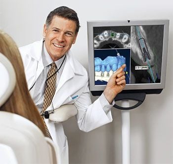
Cone beam CT imaging provides dentists with a three-dimensional view of mouth, jaw, teeth, and nasal cavity. These images contain invaluable clinical information and help reduce the need for invasive procedures, shorten treatment time, and make treatment plans more effective and efficient.
With 3D scans, dentists and dental specialists can now evaluate:
- Soft tissue size and location
This is especially important in diagnosing and treating sleep apnea, where soft tissue might be blocking the airways during sleep. These images will also show tumors and irregular growths, and can help your dentist plan for oral surgery.
- Location of teeth, including impacted teeth or teeth that haven’t grown in yet
Knowing the exact location and size of your teeth helps in planning treatment for braces or impacted teeth.
- Location, size and density of jaw bone
Knowing the location, size, density of your bones will help determine the best plan of action to take if you need an implant or jaw surgery.
When you get a cone beam CT scan, an imaging device rotates around your head. The scanner records between 150 and 600 different X-ray views in under a minute and sends the scans to a computer where a virtual, three-dimensional model is created from the images. The model can be rotated from side to side or up and down, magnified, or viewed from any angle needed. So not only can your dentist see your entire tooth’s anatomy, they can zoom in to see the condition of your root canal itself. This allows your dentist or dental specialist to prepare your procedure, or examine your health in great detail without you having to sit in an uncomfortable position, or without you even needing to be present.
Like X-rays, CT scans are associated with low amounts of radiation exposure, so it’s important to consider the risk before getting a scan. Most often, the benefits of getting a CT scan outweigh the risks, but it’s particularly important to be cautious for those with preexisting health conditions.